
 By Lina Hope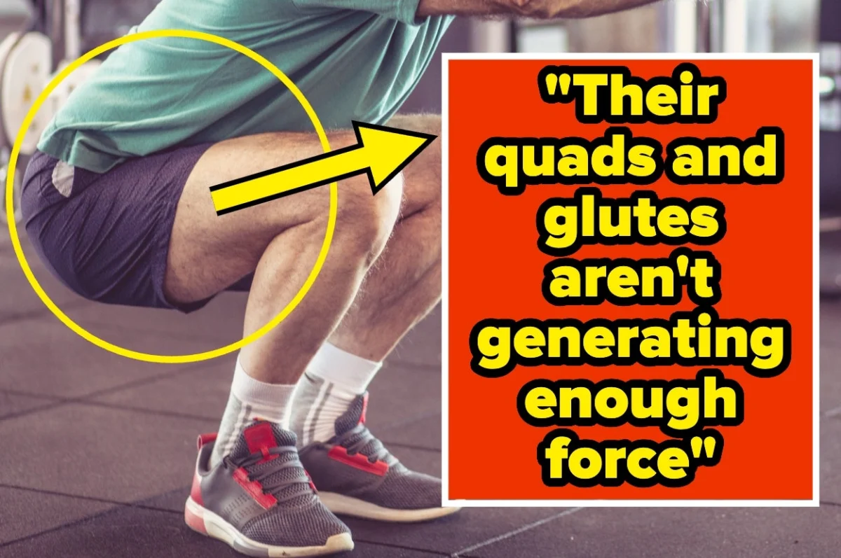
October 3, 2026 — When society evaluates the physical trajectory of aging, the metrics are usually superficial. People scan mirrors for the silver threads of gray hair, trace the emergence of crow’s feet around the eyes, or note a general slowing of pace during a brisk morning walk. These are the visible, cultural markers of getting older.
However, orthopedic specialists, physical therapists, and geriatric mobility researchers look elsewhere entirely when assessing functional longevity. They turn their attention to a part of the anatomy rarely discussed in polite society, yet central to human survival and autonomy: the backside. Specifically, they examine the gluteal muscles.
"The legs are the engine of independence," explains Scott McAfee, an orthopedic physical therapist with Movement-X. "They drive you up out of a chair, help stabilize you when your weight shifts, and help you stay upright as you move."
As research into aging and physical independence accelerates, medical professionals are shifting away from cosmetic metrics of time passing. Instead, they are focusing on structural metrics—how well the body’s primary power generators hold up against gravity, and what happens when those muscles quietly slip into decline.
Main Facts: The Decline of the Gluteal Engine
For decades, the medical community has understood that muscle mass diminishes with age—a process known as sarcopenia. Yet, recent clinical observations and historical studies highlight that this loss is rarely uniform. The lower body, and the gluteal complex in particular, bears the brunt of a sedentary lifestyle and the natural physiological shifts of getting older.
A landmark 2014 study published in BMC Geriatrics provided stark empirical backing for this observation. Researchers compared 58 adults over the age of 65 who had experienced a fall within the previous year against a control group of peers who had remained fall-free. While muscle composition changes were evident across the entire lower body, the gluteal region bore the most dramatic alterations.
Most notably, the magnetic resonance imaging and body composition data revealed that the amount of intramuscular fat within the gluteus maximus was twice as high in the group of recent fallers compared to those who had not fallen. Researchers concluded that fatty infiltration and muscle degradation in the gluteal region directly undermine core stability, impair balance, and significantly elevate the risk of sudden, dangerous falls.
This degradation does not happen overnight. It is a gradual erosion of strength that often goes unnoticed until an individual is forced into a high-stakes physical situation—such as catching themselves on an icy patch, stepping off an uneven curb, or simply trying to rise from a deep sofa without assistance.
Chronology: How Specialists Spot Hidden Vulnerabilities
Mobility experts do not wait for a catastrophic fall to assess a patient’s risk profile. Instead, they use a series of clinical evaluations that break down daily human movement into its fundamental components: rising, descending, stabilizing, and turning.
Phase 1: The Ascent and Descent
The assessment often begins with a deceptively simple metric: the 30-second chair stand. Patients cross their arms over their chest and rise to a full standing position, then sit back down as many times as possible within half a minute.
According to McAfee, the quantitative count matters just as much as the qualitative execution. "A fit 62-year-old woman knocking out 18 is in great shape," McAfee notes. "The same woman managing only 9 or 10 is telling me her fall risk may be climbing."
Equally revealing is how the movement is performed. If a patient must push off their thighs or rely heavily on armrests to generate upward momentum, clinicians immediately flag the individual as high-risk.
The descent is scrutinized with equal intensity. "A strong mover lowers under control, while a rising fall risk drops the last few inches into the seat because the muscles that decelerate the body are already failing," McAfee explains. "I watch the descent as closely as the ascent."
Phase 2: Torso Compensation and Asymmetry
As patients rise, specialists examine the kinetic chain for compensatory movements. Priyank Patel, a Brooklyn-based chiropractor who treats older adults for hip, low-back, and gait abnormalities, watches the trunk and hips closely during the transition from sitting to standing.
"If they pitch their torso forward a lot to get momentum going, that tells me their quads and glutes aren’t generating enough force on their own, so the body is borrowing momentum instead," Patel says.
Asymmetry provides another critical diagnostic clue. A minor structural imbalance—such as one hip hiking up prematurely during an ascent or a knee wobbling inward—serves as an immediate red flag for underlying weakness or neural control issues.
Phase 3: Dynamic Floor Transitions
Getting out of a chair is only part of the puzzle. Austin Misiura, an orthopedic physical therapist at Pure PT Miami, utilizes a more demanding test: getting down to the floor and returning to a standing position completely unassisted by hands or furniture, paired with single-leg balance trials.
Misiura argues that floor-rise tests capture vulnerabilities that traditional chair assessments miss entirely. "For the chair stand, there’s almost no mobility requirement," Misiura observes. "The mobility component of the floor stand is completely missed by pretty much every other standardized test, as essentially everyone can be in a standing or sitting position with little to no insight on mobility."
Furthermore, Misiura notes that patients often mask their physical decline during routine walking tests because they modify their gait in a clinical setting. The true deficit emerges when concealment is impossible. "Clients won’t even want to get on the floor, or they will need help right away," he says. On their feet, these same individuals exhibit a distinct "shuffling gait" designed to secure contact with the ground as quickly as possible.
Phase 4: The Rotational Load Test
Turning represents another high-risk biomechanical event. Dr. Alexander Acosta, a physician and medical consultant for SonderCare, points out that changing direction places an asymmetric, dynamic load on a single leg.
"A turn is a move that causes someone to place load on one leg for a moment, revealing balance and strength," Dr. Acosta says. "A sicker patient will not be able to disguise this."
Dr. Acosta measures functional capacity by counting the steps required for a patient to complete a full 360-degree turn. Healthy, robust adults typically accomplish this in three or four steps. "I begin to pay more attention when someone needs five steps or more, or reaches out for a wall or chair," he notes, recalling a patient who required six steps and hand support on a wall to execute a single turn. "The difference between four steps and six isn’t subtle once you know what to look for."
Supporting Data: Benchmarks for Functional Health
To help individuals gauge their own physical independence, public health agencies and clinical researchers have established standardized normative values.
The Centers for Disease Control and Prevention (CDC), through its STEADI (Stopping Elderly Accidents, Deaths, & Injuries) initiative, provides clear benchmarks for the 30-second chair stand test:
- Men (Early 60s): Should manage a minimum of 14 successful stands.
- Women (Early 60s): Should manage a minimum of 12 successful stands.
- Trend: Thresholds decrease systematically with each passing decade, reflecting expected age-related physiological declines—though maintaining rigorous physical activity can arrest or even reverse these trends.
For single-leg balance, clinical guidelines generally look for an individual’s ability to stand unassisted on one foot with eyes open for at least 10 seconds per side. Inability to maintain balance within the first few seconds strongly correlates with diminished core and gluteal stability.
Official Responses and Clinical Perspectives
The medical consensus surrounding gluteal strength marks a significant evolution in how physical therapy and preventative geriatrics intersect. Rather than treating falls as random accidents of old age, specialists view them as predictable outcomes of localized muscular neglect.
Yet, a core message from the clinical community is one of radical optimism: muscle tissue responds to progressive training at any age.
"One low score should not be discouraging," Scott McAfee emphasizes. "Maybe just a nudge to see your physical therapist to improve it."
Unlike structural dermatological changes or internal cellular shifts, skeletal muscle retains its plasticity deep into later life. When stimulated properly through resistance training, protein synthesis, and balance work, aging muscle fibers can regain volume, neural recruitment, and functional output.
Implications: A DIY Assessment and the Path Forward
For adults looking to evaluate their physical resilience before a minor weakness turns into a life-altering fall, mobility experts recommend a straightforward, self-administered screening protocol.
How to Test Your Glutes at Home
Note: Always ensure a stable support surface or a helping partner is nearby. Stop immediately if you experience dizziness, joint pain, or unsteadiness.
- The 30-Second Chair Stand: Set a timer for 30 seconds. Cross your arms over your chest and complete as many full sit-to-stands as safely possible. Compare your score against CDC age-adjusted norms, noting whether you require arm assistance or lose control on the descent.
- The Floor-to-Stand Test: If cleared for safe movement, lower yourself to the floor and return to a standing position without using your hands, knees against furniture, or wall supports.
- The Single-Foot Balance: Stand near a wall or counter for safety. Lift one foot and time how long you can maintain balance with your eyes open, aiming for 10 seconds per leg.
- The 360-Degree Turn: Stand safely in an open space or near a door frame. Turn completely around in a full circle while counting your steps. Healthy movement should require no more than 3 to 4 steps without grabbing structural supports.
The Takeaway
Cosmetic signs of aging—such as graying hair or fine lines—remain entirely impervious to exercise routines and physical therapy interventions. They are passive markers of time passing.
The muscles that lift a human body out of a deep armchair, rescue them from a fall on the floor, or safely guide them around a sharp corner, however, are entirely different. They are active, responsive, and ready to be rebuilt. By shifting our attention from the mirror to our movement, we reclaim agency over the physical timeline of our lives.
 Dwi Wanna
  Neng Nana
  Laily UPN