
 By Lina Irawan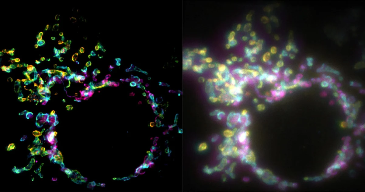
Main Facts
Cellular biology has entered a potentially transformative era with the introduction of a groundbreaking fluorescence microscopy technique called SPIFFI (spatial polarization-induced fluorescence fluctuation imaging). Developed by a team of researchers at the Laboratory of Nanoscale Biology (LBEN) within EPFL’s School of Engineering—namely Wei Guo, Lely Feletti, and Aleksandra Radenovic—the method allows scientists to capture high-contrast, super-resolution images of internal cellular structures from a single exposure.
Detailed in a prominent study published in the scientific journal Nature, SPIFFI overcomes a major historical bottleneck in optical microscopy: the trade-off between spatial resolution and temporal acquisition speed. Traditional super-resolution techniques often require the compilation of hundreds or even thousands of individual frames to construct a single high-resolution image. Because living cells are inherently dynamic—constantly shifting, dividing, and reacting—these multi-frame capture methods frequently result in motion blur, rendering real-time observation of living cellular processes extremely difficult, if not impossible.
SPIFFI solves this fundamental limitation by harnessing the polarization properties of fluorescent light. Instead of accumulating temporal data across multiple exposures, the system splits fluorescent light emitted by targeted molecules into four distinct polarization-sensitive channels. By comparing these channels simultaneously within a single frame, the system recovers fine structural details that are typically lost in conventional widefield microscopy.
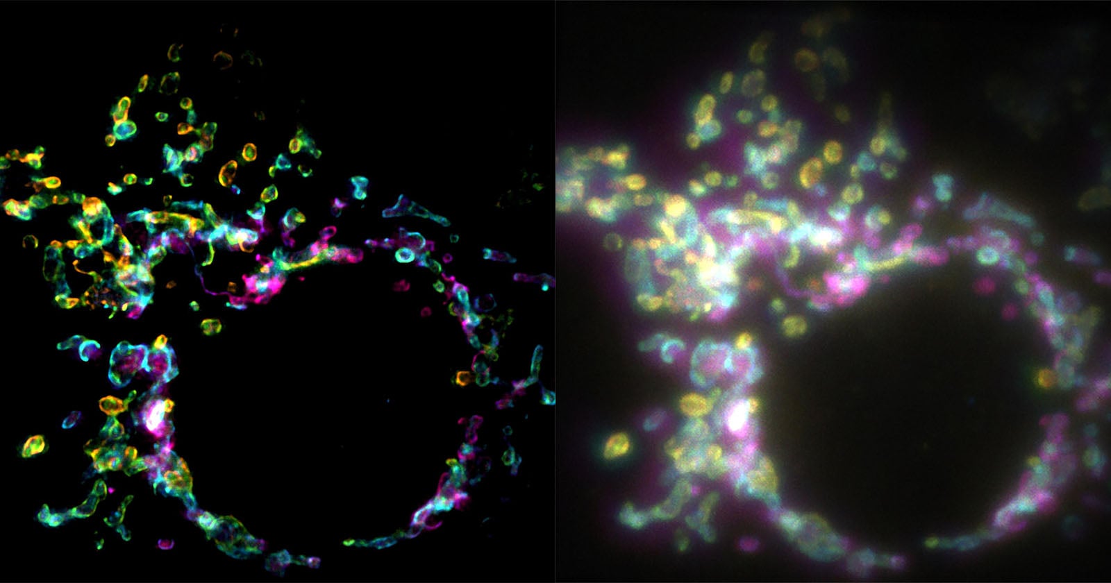
Crucially, the technology achieves a twofold improvement in resolution within a single image, resolving structures as small as 160 to 170 nanometers. Furthermore, when integrated with existing fluctuation-based post-processing methods, the effective resolution can be pushed down to approximately 80 nanometers. Perhaps most appealing to institutional laboratories, SPIFFI’s optical hardware can be retrofitted onto existing fluorescence microscopes, presenting a cost-effective and practical alternative to investing in entirely new, dedicated imaging systems.
Chronology of Development
The path to developing SPIFFI spans years of methodical research in nanoscale physics, photonics, and cellular biology at EPFL. While the formal unveiling of the technique occurred with its publication in Nature, the groundwork was laid much earlier as researchers sought to address the inherent limitations of conventional temporal fluctuation imaging.
- Early Phase – Identifying the Temporal Bottleneck: For decades, super-resolution microscopy revolutionized how biologists viewed the interior of cells, allowing science to peer past the classical diffraction limit of light. However, techniques like STORM (Stochastic Optical Reconstruction Microscopy) and PALM (Photoactivated Localization Microscopy) relied heavily on capturing sequential time-lapse data to pinpoint the exact locations of fluorescent probes. Researchers recognized that while this worked well for fixed, dead cell samples, it was fundamentally unsuited for capturing rapid, live-cell phenomena because biological structures moved faster than the data acquisition rate.
- The Conceptual Shift – Leveraging Polarization: The LBEN team shifted their focus from temporal information (how molecules change over time) to spatial polarization information (how individual fluorescent molecules are oriented in space at a single moment). Because fluorescent molecules emit light that oscillates preferentially along specific vectors depending on their spatial orientation, researchers hypothesized that these polarization signatures contained hidden structural details.
- Hardware and Algorithm Design: Over subsequent months, the team designed specialized optical components capable of splitting incoming fluorescent emissions into four polarization-sensitive channels simultaneously. Alongside the hardware, they developed computational algorithms to process these multi-channel snapshots instantly, converting complex polarization data into sharp, high-contrast structural images.
- Experimental Validation: The team tested the system on complex biological structures, successfully capturing rapid physiological events such as mitochondrial outer membrane dynamics, cellular splitting (cytokinesis), and mitochondrial fusion events with unprecedented clarity.
- The Nature Publication: The culmination of these efforts was formally documented and peer-reviewed, leading to the publication of the SPIFFI paper in Nature, instantly generating widespread interest within the global biophotonics community.
- Current and Future Steps: Having proven the viability of the optical hardware, the research team is currently focusing on engineering more compact iterations of the system to maximize usability and streamline integration into standard laboratory workflows.
Supporting Data and Technical Specifications
To fully understand the significance of SPIFFI, it is helpful to examine the quantitative data and optical metrics associated with the technology as reported by the EPFL research team:
- Resolution Enhancement: SPIFFI immediately improves standard widefield resolution by a factor of two within a single captured frame.
- Absolute Spatial Resolution: The technique natively resolves biological structures down to approximately 160 to 170 nanometers.
- Post-Processing Enhancement: When SPIFFI single-frame captures are combined with existing fluctuation-based post-processing software pipelines, the resolution is further refined to roughly 80 nanometers.
- Optical Channel Breakdown: The system splits incoming fluorescence into four distinct polarization-sensitive channels, allowing the software to analyze molecular orientation vectors concurrently.
- Dynamic Capabilities: Unlike legacy methods that demand hundreds or thousands of sequential frames for a single resolved image, SPIFFI ensures that every single frame is super-resolved, effectively opening the door to true high-definition, high-speed super-resolution video of living cells.
- Hardware Compatibility: The modular nature of the SPIFFI optical setup means it can be integrated directly into pre-existing fluorescence microscope architectures without requiring a ground-up redesign of laboratory infrastructure.
Official Responses and Expert Insights
The introduction of SPIFFI has drawn enthusiastic commentary from its creators and the broader scientific community, highlighting the paradigm shift it represents for live-cell imaging.
Aleksandra Radenovic, who leads the Laboratory of Nanoscale Biology (LBEN) in EPFL’s School of Engineering, emphasized the unprecedented observational power the technique unlocks for biologists:
"SPIFFI can capture fast-moving processes within cells, while enabling high-throughput, multi-dimensional imaging beyond the limits of conventional microscopes."
First author and LBEN PhD student Wei Guo elaborated on the distinct mechanical advantage of the new method over historical approaches, pointing out the elimination of the temporal trade-off:
"Essentially, previous approaches used temporal information to resolve spatial resolution, but this doesn’t work very well on living cells."
Guo further highlighted the transition from static photography to dynamic cinematography at the nanoscale:
"With previous techniques, taking many images would only result in one super-resolved frame. With SPIFFI, every frame is super-resolved, meaning we can now produce super-resolution videos of live cells. We also seamlessly integrated SPIFFI images with existing fluctuation-based methods for post-processing, achieving resolutions of about 80 nanometers."
Independent biophysicists and microscopists reviewing the Nature publication have similarly praised the method for its pragmatic approach. By eschewing the need for ultra-high-speed camera arrays or exotic, toxic photo-switching chemicals often required in traditional super-resolution workflows, SPIFFI lowers the barrier to entry for standard molecular biology laboratories wishing to upgrade their analytical capabilities.
Implications for Science, Medicine, and Industry
The practical implications of SPIFFI extend far beyond academic curiosity, promising to accelerate discoveries across several critical domains of life sciences and medical research:
1. Real-Time Virology and Immunology
Understanding how viruses enter cells, replicate, and evade immune responses requires tracking fast-moving viral particles and host cell membrane dynamics in real time. Because SPIFFI enables the generation of super-resolution video streams of living cells, virologists can observe host-pathogen interactions with a level of clarity previously restricted to frozen, static snapshots.
2. Neuroscience and Synaptic Transmission
Neuronal communication relies on rapid vesicle fusion and neurotransmitter release at synapses—processes that occur on millisecond timescales and nanoscale spatial dimensions. SPIFFI’s ability to resolve structures down to 80 nanometers in single frames offers neuroscientists a powerful new lens to study synaptic plasticity, neurodegenerative disease mechanisms, and intracellular transport without motion artifacts.
3. Pharmacology and Drug Discovery
Pharmaceutical research frequently utilizes live-cell assays to monitor how candidate drugs affect cellular structures, organelle health, and receptor distributions. The integration of SPIFFI into standard fluorescence microscopes means high-throughput screening facilities could potentially upgrade their existing hardware to evaluate drug efficacy at nanoscale resolution in real time, streamlining the identification of promising therapeutics.
4. Accessibility and Laboratory Economics
Perhaps one of the most immediate commercial implications is cost-efficiency. Advanced super-resolution microscopy setups often cost hundreds of thousands of dollars and require dedicated cleanroom environments or specialized operational expertise. By designing SPIFFI to be retrofittable onto existing fluorescence microscopes—and actively working on making the hardware modules more compact—the EPFL team has laid the groundwork for widespread commercial adoption. As manufacturing partners look to package these polarization-splitting modules into user-friendly commercial add-ons, high-end super-resolution imaging could soon become a standard feature in routine biological laboratories worldwide.
 Reynand Wu
  Basiran
  Muslim